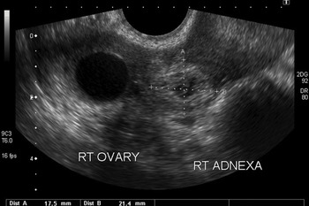
Figure 2 from Unilateral bartholin gland cyst in a pregnant heifer
By A Mystery Man Writer
Last updated 20 Sept 2024

Figure 2. The blood vessels on the cyst walls (arrows). - "Unilateral bartholin gland cyst in a pregnant heifer."

Photograph showing growth on the left side of rectum.
Surgical Management of Cystic Unilateral Bartholin's Gland in a Crossbred cow - Document - Gale Academic OneFile

Female Repro Flashcards
Basic Sciences in Gynaecology (Section 1) - The EBCOG Postgraduate

Brasil - Bartholin's gland adenoma in a Saanen goat - SciELO

Congenital and acquired pathology of ovary and tubular genital
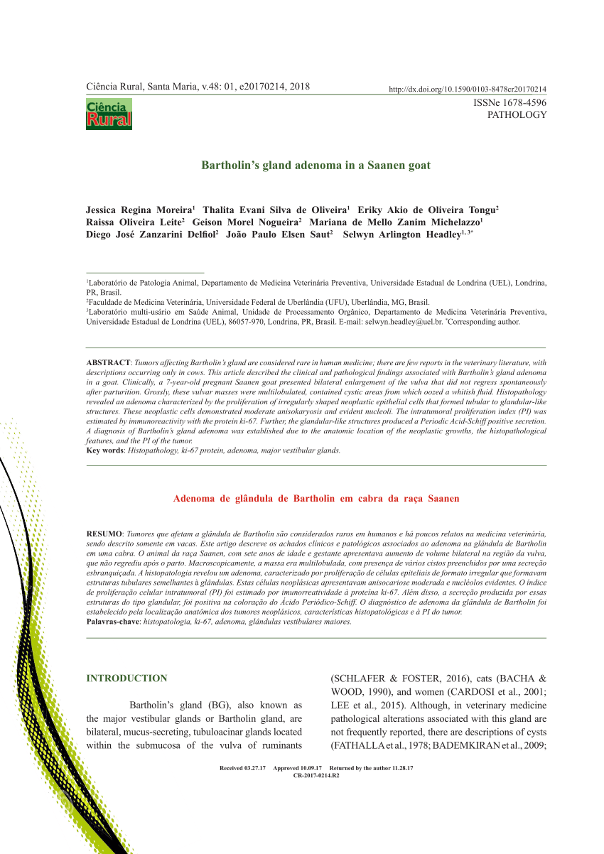
PDF) Bartholin's gland adenoma in a Saanen goat

Vaginal Vestibule - an overview

Obstetrics, PDF, Pregnancy

PDF] Bartholin Duct Cyst and Gland Abscess: Office Management

Photograph showing growth on the left side of rectum.

Figure 2 from Bilateral Massive Hematoma of Bartholin Glands after

Reproductive Anatomy and Physiology of the Nonpregnant and

LORI.bovine
Management of Bartholin's Duct Cyst and Gland Abscess
Recommended for you
 Bartholin's Cyst – Causes, Symptoms & Treatment14 Jul 2023
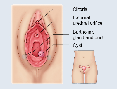 Bartholin cyst removal - Mysurgeryabroad - Medicover Hospital14 Jul 2023
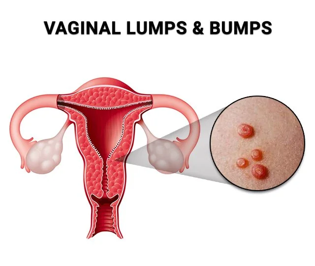 5 Possible Explanations for Vaginal Lumps & Bumps - Century14 Jul 2023
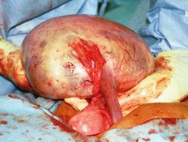 Ovarian Cysts: Practice Essentials, Background, Pathophysiology14 Jul 2023
- Is it normal for an ovarian cyst to cause bleeding? - Quora14 Jul 2023
 Ovarian cysts - Symptoms and causes - Mayo Clinic14 Jul 2023
 Atypical Presentation of a Vaginal Epithelial Inclusion Cyst14 Jul 2023
 Ovarian Cysts – Types, Symptoms, Treatment – Dr. Deepa Ganesh14 Jul 2023
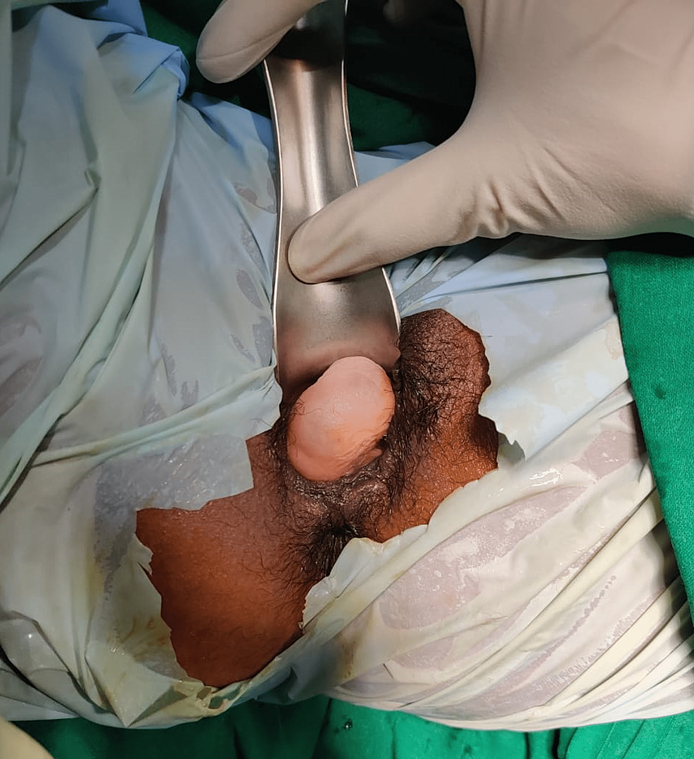 Cureus, A Rare Case of Posterior Vaginal Wall Gartner's Duct Cyst Mimicking as Genital Prolapse14 Jul 2023
 Hysteroscopy Dr. Sarah Choi - Gynaecologist and Advanced Laparoscopic Surgeon in Sydney14 Jul 2023
You may also like
 Space Odyssey Poster14 Jul 2023
 Hold Down Ceiling Tile Clips, 100 Units14 Jul 2023
 US Girls Cargo Pants Fashion Casual Sweatpants Sports Jogger Pants14 Jul 2023
- Short Push UP - Levanta Pompis GENERICO14 Jul 2023
- JORDAN SPODNIE JDN ESS LEGGING BLK PANTS DQ4448-01014 Jul 2023
/cdn.vox-cdn.com/uploads/chorus_image/image/55265151/aerie_underwear.0.jpeg) Aerie Makes the Best Mall-Brand Underwear - Racked14 Jul 2023
Aerie Makes the Best Mall-Brand Underwear - Racked14 Jul 2023 Galvanized T-Grids – Ocean International14 Jul 2023
 A Surreal Koala with a Tail of Rainbow Ribbons, Clinging To a14 Jul 2023
 DORTALA 5 Piece Dining Table and Chairs Set, Metal Frame Home Kitchen Dinette Dining Room Furniture for 4 Person, Modern Dining Table Set with Glass Table Top & 4 Leather Padded Chairs14 Jul 2023
 How to decide on yoga vs gym which is better, by Yuvaap FindYourY14 Jul 2023