Isolation, expansion and characterization of porcine urinary bladder smooth muscle cells for tissue engineering, Biological Procedures Online
By A Mystery Man Writer
Last updated 20 Sept 2024
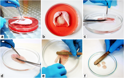
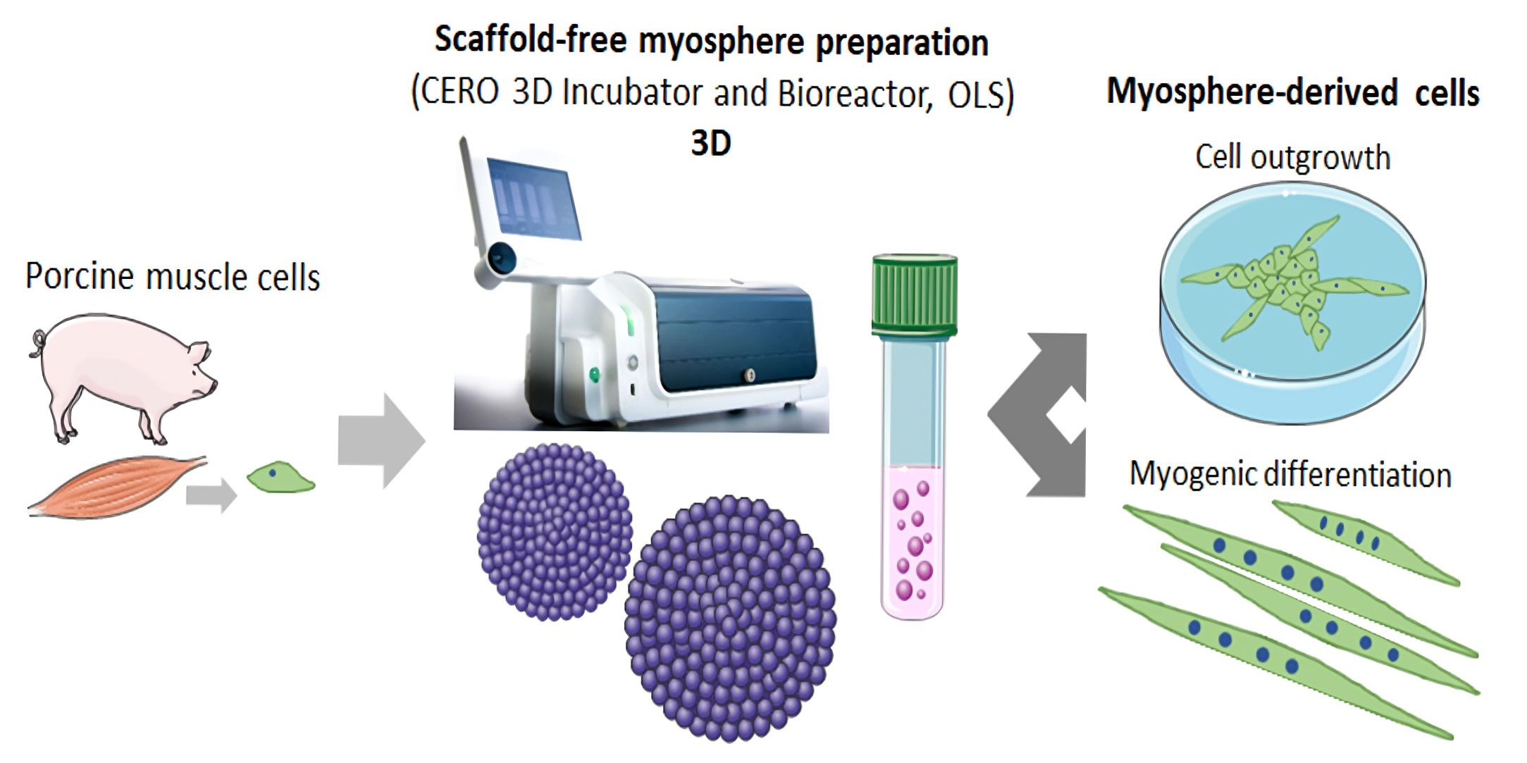
Background A key requirements for therapy utilizing the tissue engineering methodologies is use of techniques which have the capability to yield a high number of cells, from small tissue biopsy in a relatively short time. Up to date there was no optimal methods of isolation and expansion of urinary bladder smooth muscle cells (UB-SMCs). The aim of this study was to compare isolation and expansion techniques of UB-SMCs to select the most repeatable and efficient one. Method Five protocols of porcine UB- SMCs isolation including enzymatic and explant techniques and three expansion techniques were compared. Isolation effectiveness was evaluated using trypan blue assay. Cell phenotype was confirmed by immunofluorescence staining. Proliferation rate was analyzed using MTT and X- Celligence system. Cellular senescence was assessed measuring β-galactosidase activity. Results Enzymatic methods using collagenase with dispase (method I) or collagenase only (method III) allowed to isolate much larger number of cells than the methods using trypsin with collagenase (method II) and collagenase after digestion with trypsin (method IV). The success rate of establishment of primary culture was the highest when the isolated cells were cultured in the Smooth muscle Growth Medium-2 (SmGM-2). Expression of the smooth muscle markers- alpha smooth muscle actin and smoothelin was the highest for cells isolated by enzymatic method I and cultured in SmGM-2. There was no significant signs of cell senescence until the 8th passage. Conclusion The most efficient method of establishment of porcine UB-SMCs culture is enzymatic digestion of urinary bladder tissue with collagenase and dispase and culture of isolated cells in SmGM-2. This method was up to 10 times more efficient than other methods used for isolation and culture of UB-SMCs. This is an easy and consistent method for obtaining high numbers of urinary bladder smooth muscle cells.
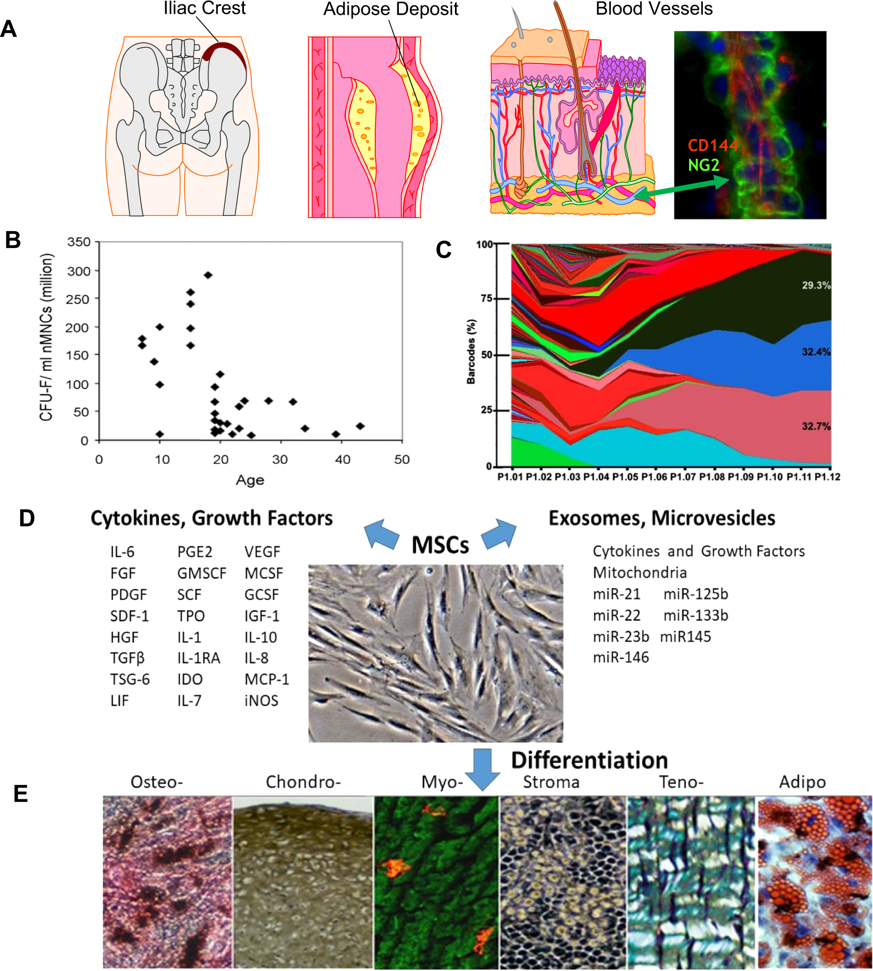
Mesenchymal stem cell perspective: cell biology to clinical progress
IJMS, Free Full-Text

Isolation, expansion and characterization of porcine urinary bladder smooth muscle cells for tissue engineering, Biological Procedures Online

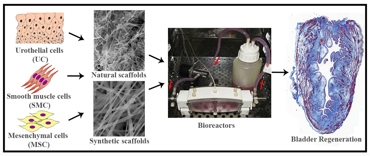
Urinary bladder and urethral tissue engineering, and 3D bioprinting approaches for urological reconstruction

Decellularized extracellular matrix biomaterials for regenerative therapies: Advances, challenges and clinical prospects - ScienceDirect
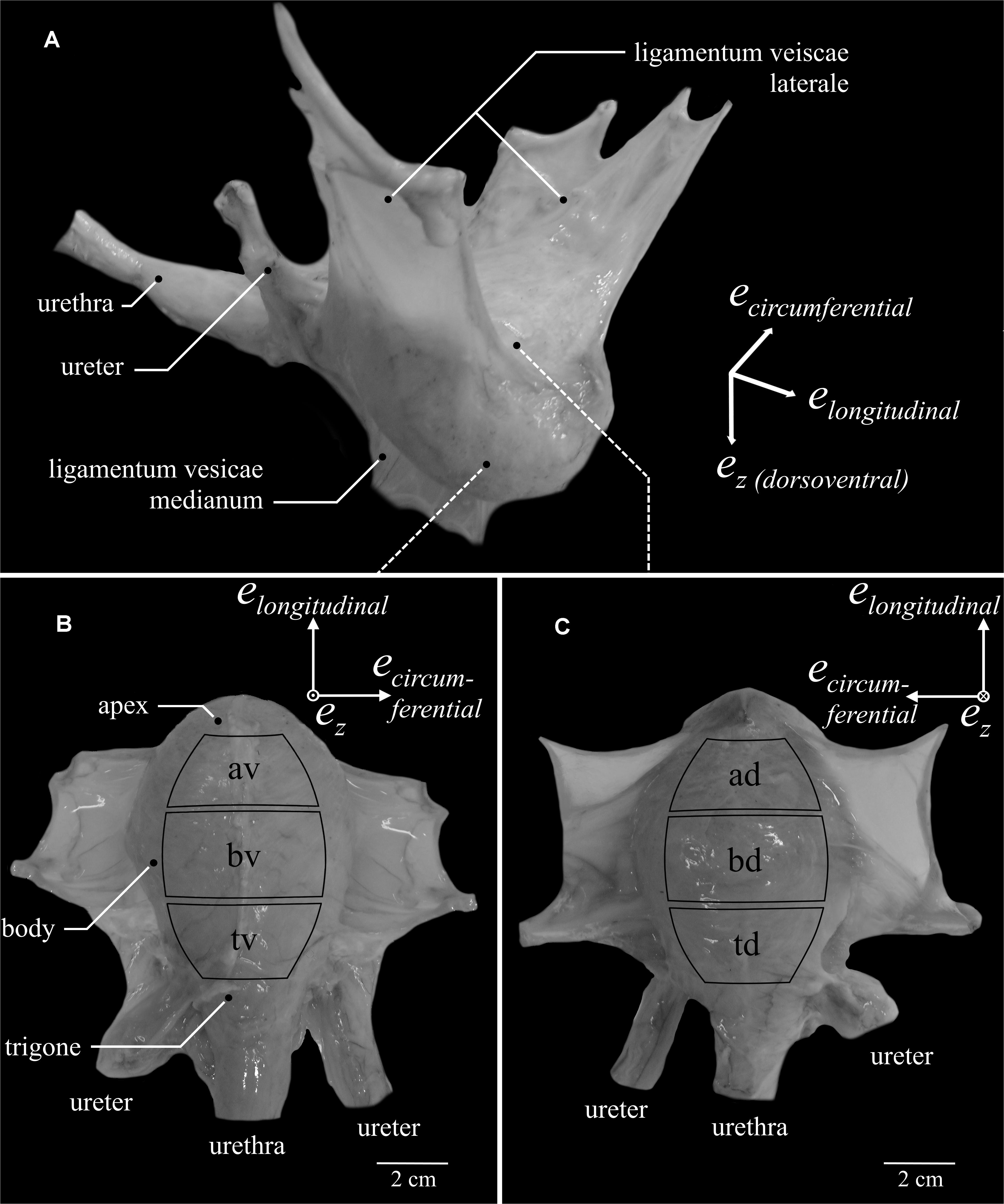
Frontiers Locational and Directional Dependencies of Smooth Muscle Properties in Pig Urinary Bladder
Isolation, expansion and characterization of porcine urinary bladder smooth muscle cells for tissue engineering, Biological Procedures Online
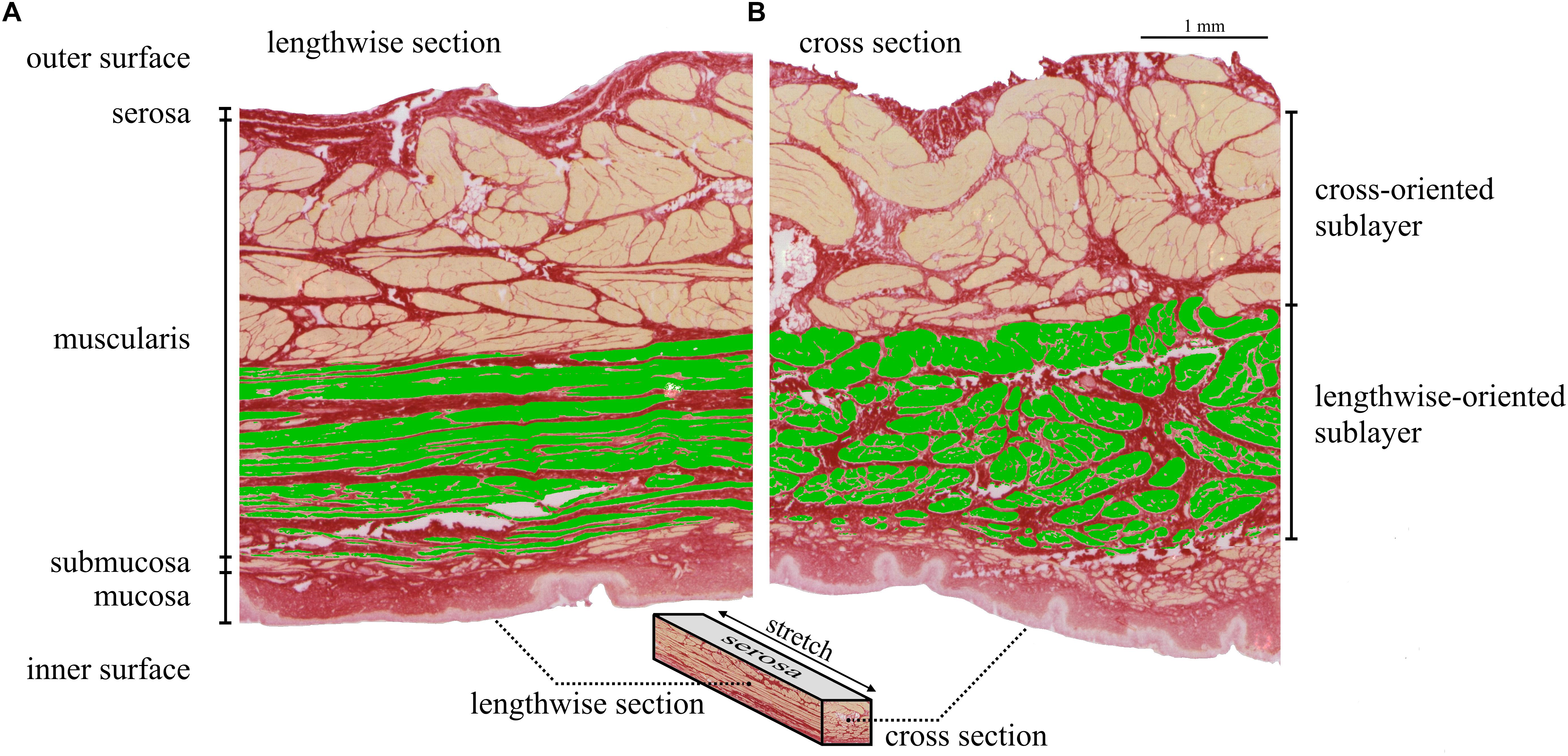
Frontiers Locational and Directional Dependencies of Smooth Muscle Properties in Pig Urinary Bladder

Isolation, expansion and characterization of porcine urinary bladder smooth muscle cells for tissue engineering, Biological Procedures Online

An examination of regenerative medicine-based strategies for the urinary bladder

Isolation, expansion and characterization of porcine urinary bladder smooth muscle cells for tissue engineering, Biological Procedures Online
Cells, Free Full-Text
Recommended for you
 Overview of Voiding - Genitourinary Disorders - MSD Manual Professional Edition14 Jul 2023
 Wearever 3-Pack Women's Black Smooth and Silky Seamless High Leg - Light Absorbency (0.25 Cup) - Incontinence Panties X-Small/Small (Fits Hip Sizes: 32-36) : Health & Household14 Jul 2023
 Corn Silk - Interstitial Cystitis, IC, Overactive Bladder, Kidney Inflammation14 Jul 2023
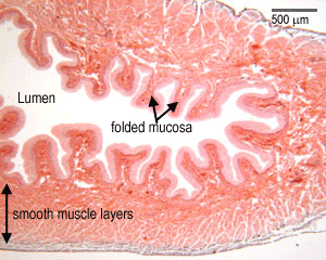 Urinary system: The Histology Guide14 Jul 2023
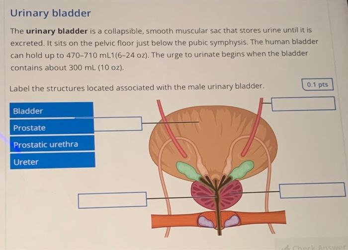
- Solved Urinary bladder The urinary bladder is a collapsible14 Jul 2023
 TotalDry® Bladder Control Pads (SP1562), 180/CS - Secure Personal Care Products SP1562 CS - Betty Mills14 Jul 2023
 Bladder Cleanse, Herbs, Ema's Herbs, Downtown Ventura14 Jul 2023
- Histological presentation of bladder smooth muscle preparations from14 Jul 2023
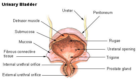 SEER Training: Urinary Bladder14 Jul 2023
- Successful bladder repair using silk fibroid14 Jul 2023
You may also like
 Analysts: Lululemon's Peloton Deal Might Not Be a Game Changer14 Jul 2023
 Women's Low Impact Strappy Sports Bra - Low Cut Wirefree Padded14 Jul 2023
 Women's sculpting uplift bra, fashion deep cup shapewear no underwire comfortable size 34-4014 Jul 2023
 Women Sexy Casual 2 in 1 Built-in Bra Nightgowns Skirt Nightdresses Soft Petticoat Nightgown,Purple-Medium : : Clothing, Shoes & Accessories14 Jul 2023
 Handbells Wesley-Knox United Church14 Jul 2023
- Diamond Underwear Body Chain Sexy Nightclub Rhinestone Chain Bra14 Jul 2023
 Paw Patrol Training Pants for Boys : : Baby14 Jul 2023
 Stradivarius Seamless faded-effect crop top - 364728734-43014 Jul 2023
- Wacoal India - Your reviews got us smiling like 😁😁😁 Get your14 Jul 2023
 Susie Delicate Blue Casual Padded Wired T-Shirt Bra14 Jul 2023