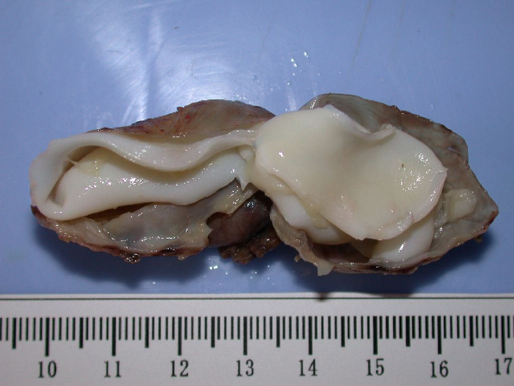
A. Cystic content was haematic. B: Hydatid membranes have the color red
By A Mystery Man Writer
Last updated 23 Sept 2024

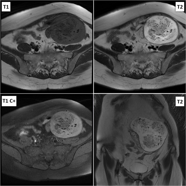
Hydatid Disease: A Pictorial Review of Uncommon Locations
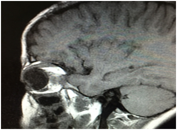
Fronto orbital approach for primary orbital hydatid cyst: Case report - MedCrave online

PDF) Giant Endocardial Blood Cyst in the Right Atrium: Echocardiographic and Magnetic Resonance Imaging Features

Cystic liver lesions: a pictorial review, Insights into Imaging

Aneurysmal Bone Cyst
Hydatid Disease: A Pictorial Review of Uncommon Locations
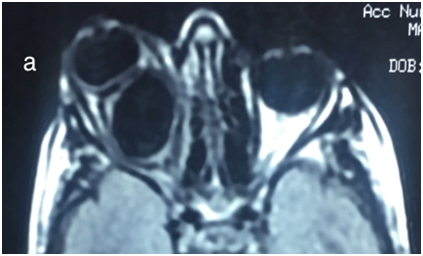
Fronto orbital approach for primary orbital hydatid cyst: Case report - MedCrave online

A. Cystic content was haematic. B: Hydatid membranes have the color red
Hydatid disease, Radiology Reference Article

Cardiac MRI images. A, Steady-state free precession sequence showing a
Primary Hydatid Cyst: An Unusual Cause of a Mass, Iynen

PDF) Stroke-Associating Acute Limb Ischemia Due to the Rupture of a Hydatid Cyst

Primary subcutaneous hydatid cysts: A review of 22 cases - ScienceDirect
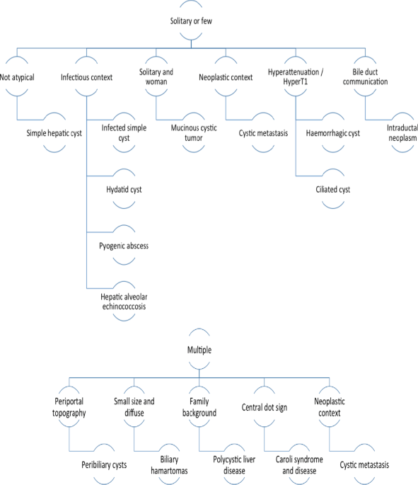
Cystic liver lesions: a pictorial review, Insights into Imaging
Recommended for you
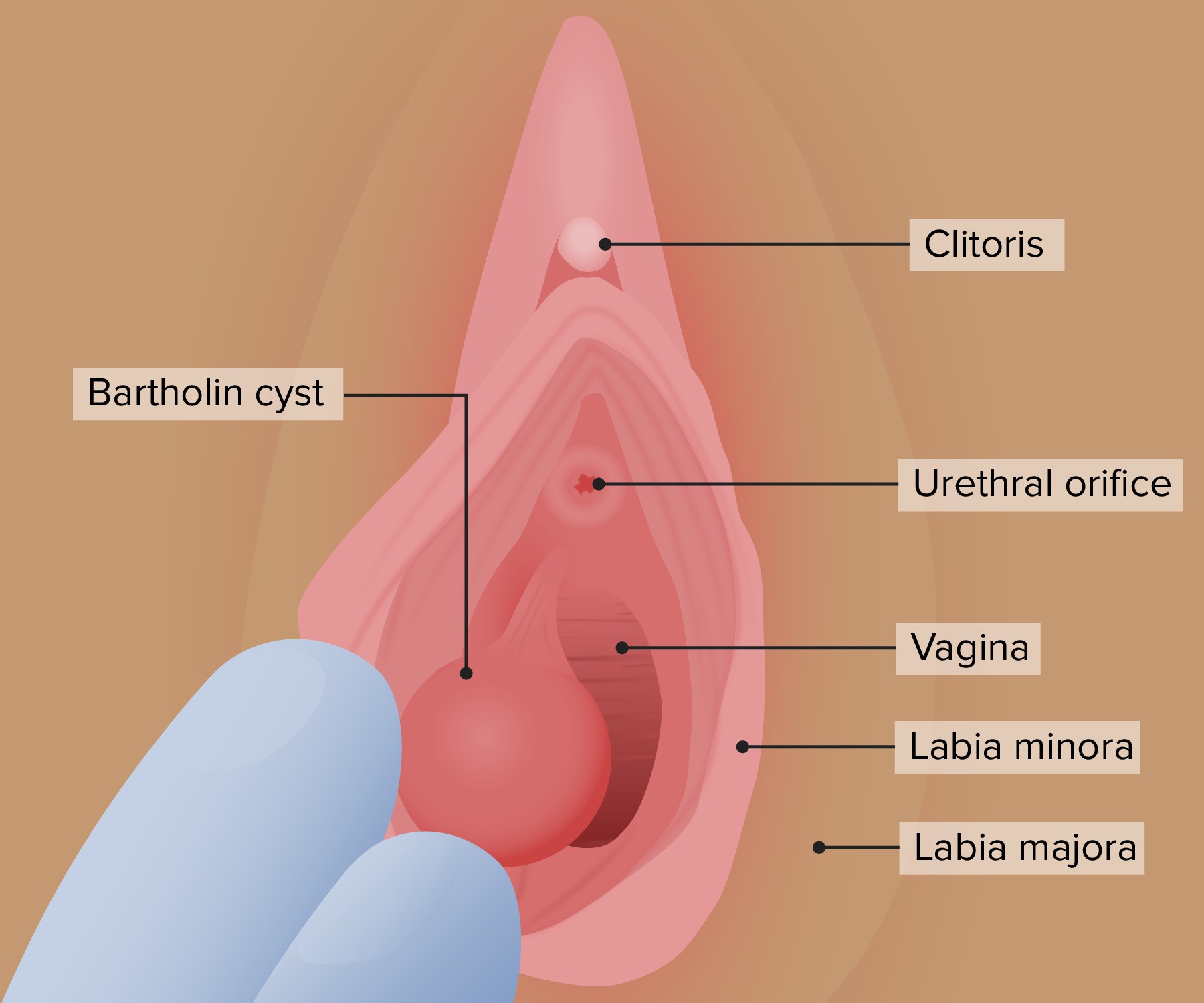 Benign Vulvar Conditions Concise Medical Knowledge14 Jul 2023
 Contents of Bartholin cyst rupture 9/10 Satisfied : r/popping14 Jul 2023
 Angiomyofibroblastoma: A rare benign gynecologic tumor mistaken14 Jul 2023
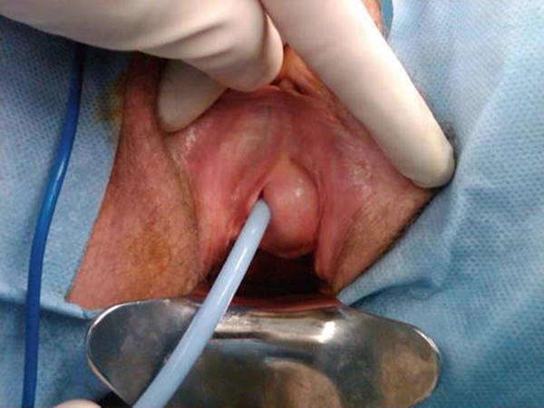 Excision of Vaginal Cysts - Female Pelvic Surgery14 Jul 2023
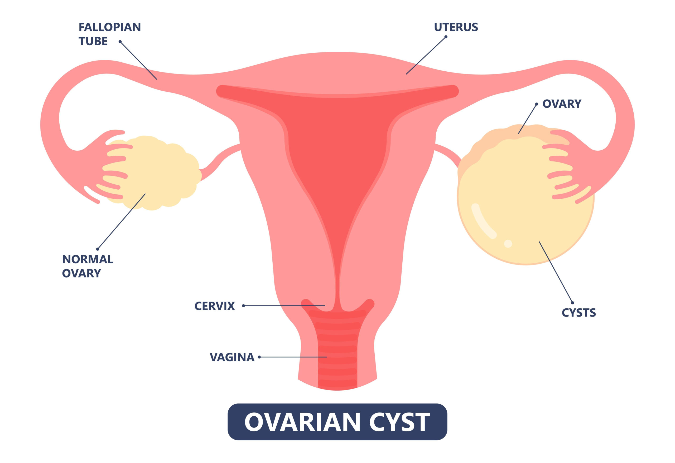 Everything You Need to Know About Ovarian Cysts: Causes, Diagnosis14 Jul 2023
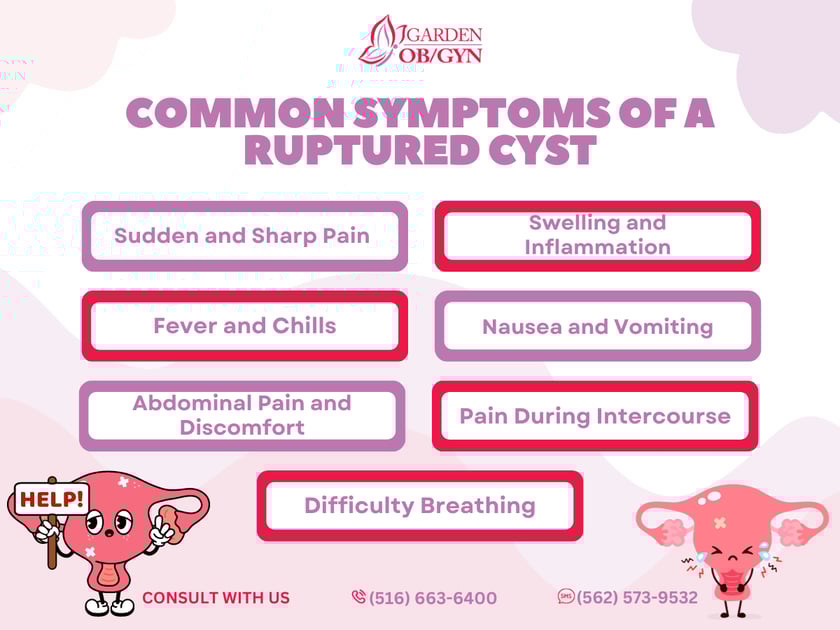 Common Symptoms of a Ruptured Cyst: Garden OBGYN: Obstetrics14 Jul 2023
 Posterior vaginal wall Gartner's duct cyst. - Abstract - Europe PMC14 Jul 2023
 Ruptured corpus luteal cyst of pregnancy with massive hemoperitoneum at14 Jul 2023
 Vaginal cysts: An important differential diagnosis in the anterior compartment - ScienceDirect14 Jul 2023
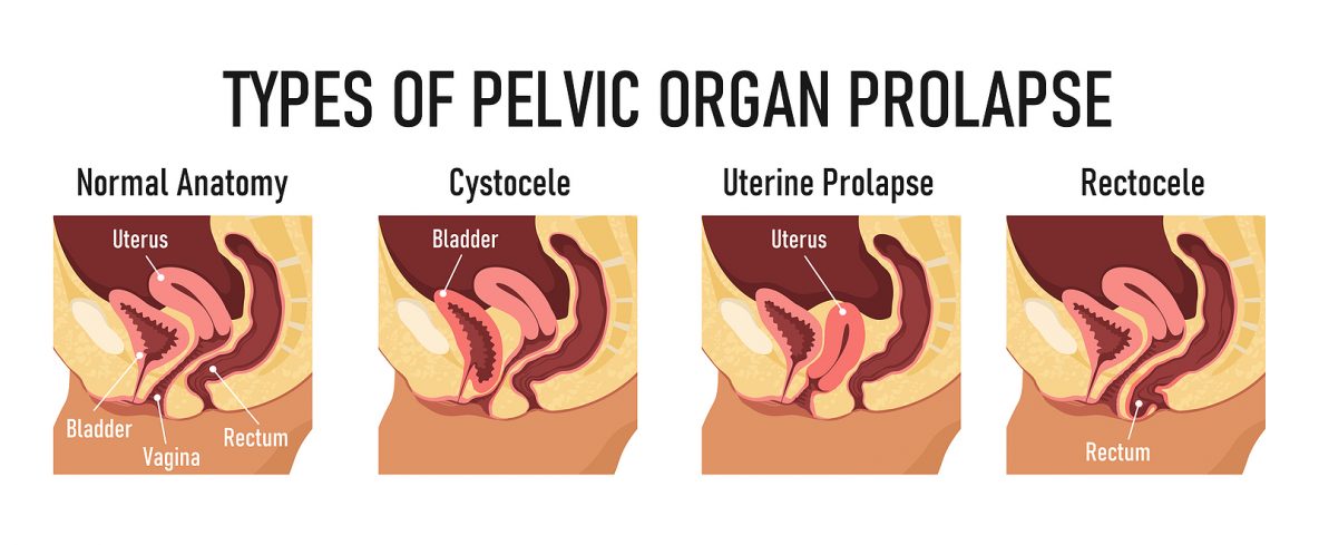 The Major Types of Pelvic Organ Prolapse and Their Differences14 Jul 2023
You may also like
 Logitech Harmony 650 Infrared All in One Remote Control, Universal Remote Logitech, Programmable Remote (Silver)-915-00015914 Jul 2023
 Accesorios y Ropa para Niñas - 2-14 años - vertbaudet14 Jul 2023
 Under Armour Women's 6 Pack Breathe Lite Ultra Low Socks14 Jul 2023
 11 Amazing Benefits Of Resistance Bands14 Jul 2023
 Dark Forest LAC longline with Align HR crop 23” : r/lululemon14 Jul 2023
 Um dos melhores surfistas do mundo está retido no aeroporto de14 Jul 2023
 Sport people woman and man flat fitness activities14 Jul 2023
 lululemon athletica, Tops14 Jul 2023
 Black Bodysuit - Satin bodysuit - Lace Bodysuit - Cami Bodysuit - Lulus14 Jul 2023
 80s Colorful Graffiti Funny Sweatpants – D&F Clothing14 Jul 2023